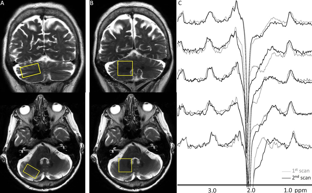
Figure 1.
Representative right cerebellar cortex (A) and dentate (B) voxel placements on T2-weighted coronal and axial slices. Short TE data were obtained from both VOIs, whereas GABA spectra were only obtained from the dentate VOIs. (C) shows pairs of repeated right cerebellar dentate GABA-edited difference spectra. Good visual correspondence was observed between the repeated measurements.

