
Fig. 2.
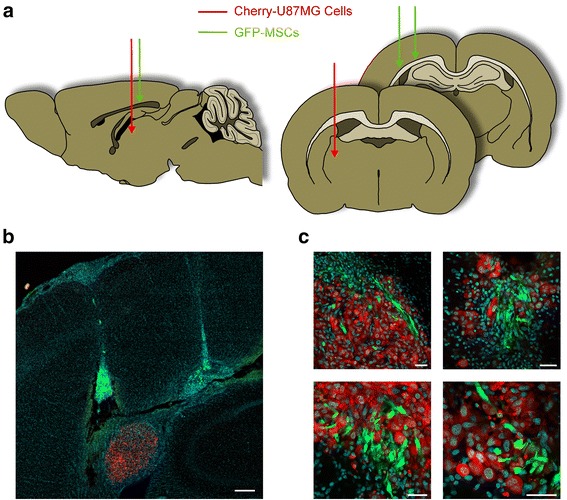
Orthotopic brain tumor xenograft. Cherry-U87MG cells and GFP-MSCs, either unloaded or loaded with PTX, were grafted in the right striatum of immunosuppressed rats. The two cell lines were injected into distinct brain sites (a). Low-power picture of a coronal section through the injection sites of GFP-MSCs showing their migration towards the Cherry-U87MG cells. Scale bar = 250 μm (b). High-power pictures showing the red tumor that appears to be massively colonized by the green MSCs. Scale bars = 40 μm (c). GFP green fluorescent protein, MSC mesenchymal stem/stromal cell