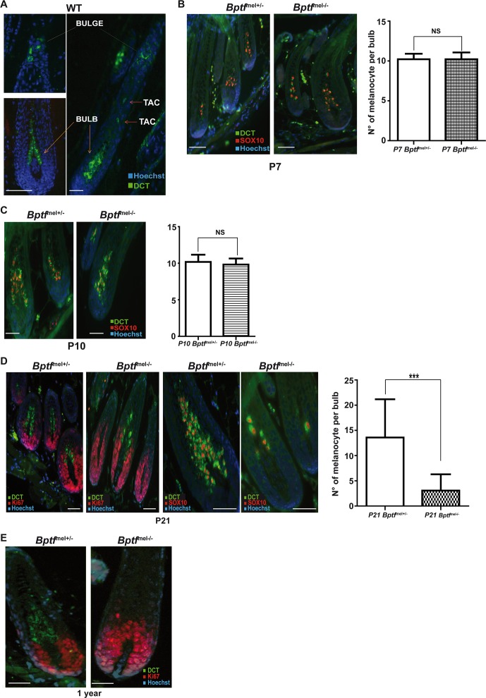
Fig 6. Loss of differentiated melanocytes in the bulb region of post-natal Bptf mel-/- mice.
A. Staining of a dorsal wild-type hair follicle with antibody against Dct showing the presence of Dct-labelled cells in the bulge corresponding to MSCs, in the shaft corresponding to TACs and in the bulb corresponding to differentiated melanocytes. B-C. Staining of dorsal hair follicles from mice with the indicated genotypes with antibodies against Dct and Sox10 to detect differentiated melanocytes in the bulb. Quantification is shown on the right. N = 30. D. Staining of dorsal hair follicles from mice with the indicated genotypes with antibodies against Dct and Ki67 to detect melanocytes in the bulb of 21 day-old animals. Quantification is shown on the right. N = 50. E. Staining of dorsal hair follicles from one year-old mice with antibodies against Dct and Ki67 illustrating the absence of melanocytes in the bulb of Bptf-mutant animals. Scale bars represent 50μm. NS = Non significant. ***P < 0.001

