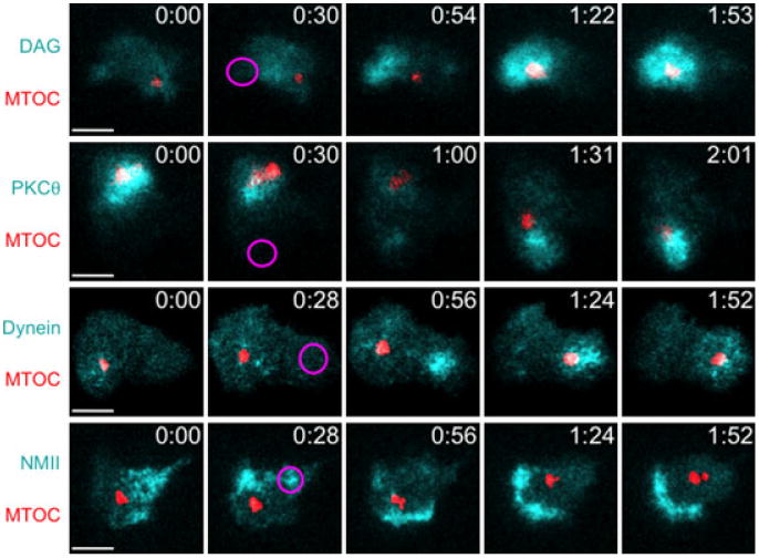
Fig. 2. Analysis of microtubule-organizing center (MTOC) polarization by T-cell antigen receptor photoactivation.

The 5C.C7 T-cell blasts expressing fluorescent reporters for the MTOC (red) and the indicated signaling molecules and motor proteins (cyan) were imaged and ultraviolet (UV)-irradiated on glass coverslips containing photoactivatable peptide-major histocompatibility complex. Representative time-lapse montages are shown (time in m:ss in the upper right corner of each image) with the time and region of UV irradiation indicated by a magenta oval. Scale bars = 5 μm.
