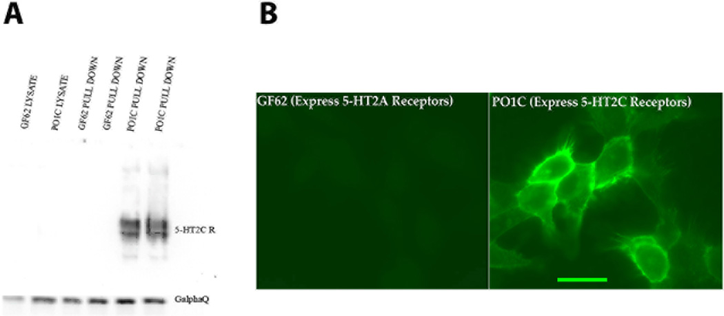
Fig1. Western blot and immunofluorescent microscopy (see Experimental Procedures) indicate that the D-12 5-HT2CR antibody used throughout this study is specific for the 5-HT2CR.
POIC cells that express 5-HT2CRs consistently showed D-12 5-HT2CR-antibody expression under both Western Blot (see A, black bars in histogram) and Immunohistochemical assessment (see B, confocal microscopy image of green cellular 5-HT2CR immunofluorescence). However, GF62 cells that only express 5-HT2AR s showed no D-12 antibody expression in either test (see A and B). Scale bar = 20 µm.

