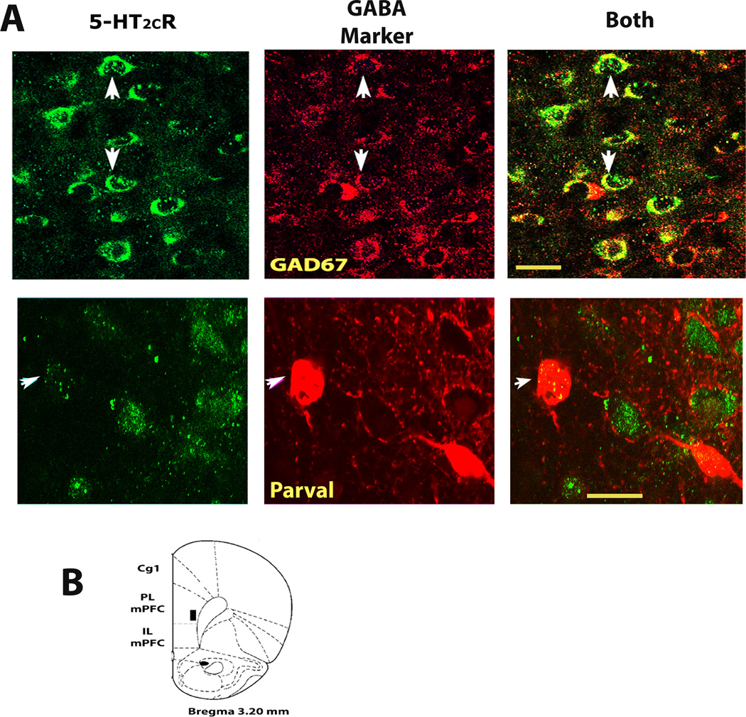
Fig2. GABA cells within the deep layers of the rat prelimbic medial prefrontal cortex (mPFC) express 5-HT2CRs.
A, confocal photomicrographs of mPFC tissue showing D-12 5-HT2CR antibody immunoreactivity (green florescence) and of cells expressing the GABA cell markers, GAD67 or parvalbumin (red immunostaining, top and bottom rows respectively). White arrows depict the identical cell across each row of photos. The left and middle photos show each antibody separately. Yellow staining in far right photos (under BOTH) illustrates 5-HT2CR and GABA cell co-localization. B, Cartoon representation of prelimbic mPFC region assessed in this experiment (see black box) according to the rat brain atlas of Paxinos and Watson (2007). 5-HT2CR, serotonin 2C receptor; GAD67, glutamic acid decarboxylase isoform 67; Parval, parvalbumin; PL mPFC, prelimbic subregion of medial prefrontal cortex. All confocal photomicrographs that were used to assess dual-immunolabeling in this report, including those presented in this figure through Fig 5, were of single optical sections (see experimental procedures, section 2.4). Scale bar = 20 µm.

