
Fig. 5.
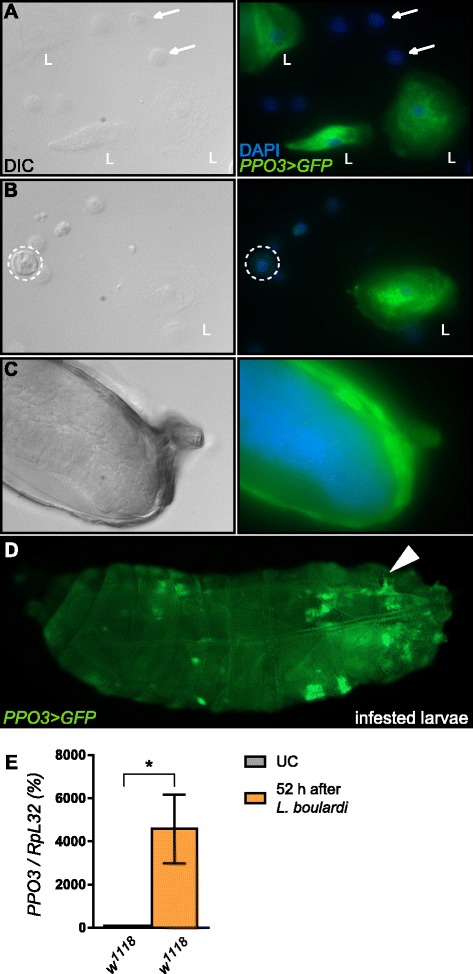
PPO3 is specifically expressed in lamellocytes. Differential interference contrast (DIC) and GFP fluorescence micrographs of hemocytes from larvae infested with Leptopilina boulardi expressing a PPO3-Gal4,UAS-GFP construct show that PPO3 is specifically expressed in all lamellocytes (L) either in circulation (a, b) or in the capsule surrounding the wasp egg (c). Plasmatocytes (arrows) and crystal cells (dashed line) do not express the reporter gene. GFP expression is green and DAPI staining is shown in blue. An overview of the whole infested larvae is shown in (d). The arrow indicates the site of the wasp larvae. Note that lamellocytes are found both around the egg and in circulation. e Quantitative reverse-transcription PCR (qRT-PCR) shows that PPO3 gene expression is higher in larvae infested with L. boulardi compared to unchallenged larvae. In the graph, 100 % corresponds to PPO3 expression levels of naive larvae. DAPI 4′,6-diamidino-2-phenylindole, GFP green fluorescent protein, UC unchallenged