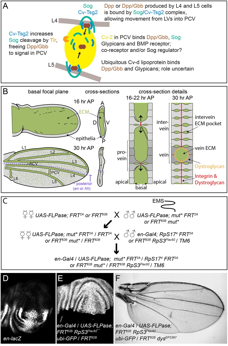
Fig 1. PCV development and the genetic screen.
(A) Model of signaling in the PCV. BMPs (Dpp and Gbb) secreted by adjacent LV cells bind to the Sog/Cv-Tsg2 complex, allowing movement into the PCV region. Cv-Tsg2 helps stimulate cleavage of Sog by the Tlr protease, freeing BMPs for signaling. Cv-2, largely bound to cells by glypicans, also binds BMPs, BMP receptors and Sog, locally transferring BMPs from Sog to the receptor complex. Cv-d also binds glypicans and BMPs and increases signaling by an unknown mechanism. (B) Diagram of vein and ECM development during the period of PCV formation in low magnification and high magnification cross-sections. As the dorsal and ventral epithelia reattach, the basal ECM of the early wing is remodeled, coming to lie in the vein channels and in scattered basolateral pockets between cells. Integrins and Dystroglycan concentrate at sites of basal-to-basal cell adhesion (both) and lining the veins (Dystroglycan). (C) Crossing scheme used to generate large homozygous mutant clones throughout the posterior compartment of the developing wing in heterozygous flies. UAS-FLPase; FRT 2A (3L) or UAS-FLPase; FRT 82B (3R) males were fed EMS and backcrossed to virgin females of the same genotype. mut* represents EMS-mutagenized chromosome. Virgin female F1 progeny were then crossed to en-Gal4; FRT 82B, RpS3 Plac92 Ubi-GFP / TM6 or en-Gal4; hs-GFP RpS17 4 FRT2A /TM6 males. Approximately 50 females were used for each of the first two crosses. (D) en expression in posterior of late third wing discs, shown using the en-lacZ enhancer trap. (E) Large posterior homozygous clones, marked by the absence of GFP (white), induced in late third instar wing disc using en-Gal4/UAS-FLPase; FRT 82B /FRT 82B RpS3 Plac92 ubi-GFP. (F) Test of the mosaic method using en-Gal4/UAS-FLPase; FRT 82B RpS3 Plac92 ubi-GFP/FRT 82B dystrophin EP3397; the adult wing shows the “detached” crossvein phenotype typical of dystrophin (dys) loss.

