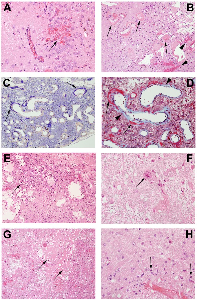
Fig 5. Pathological features in post-irradiation mouse brain.
A. Micro-hemorrhage and dilated vessels (arrow, 20X); B. Hemorrhage (arrowheads) and fibrinoid vascular necrosis (arrows) (H&E staining, 20X); C. PTAH staining shows fibrinoid vascular necrosis in dark blue (arrows, 20X); D. Trichrome staining demonstrates fibrinoid vascular necrosis (red, arrows) and collagen deposition (light blue, arrow heads)(60X); E. Macrophages surrounding damaged tissues (arrow, 20x); F. Cellular atypia (arrows, 60X); G. Edema (arrows, 20X); H. Neuronal necrosis (arrows, 60X).

