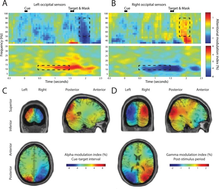
Fig 2. Time-frequency analysis and source reconstructions of attentional modulation of anticipatory alpha and stimulus-induced gamma oscillations.
(A,B) For left and right occipital MEG sensors. “Attention left” trials were compared to “attention right” trials. Bilateral attentional modulation is clearly visible in the alpha band during the cue-target interval, and bilateral modulation of stimulus-induced gamma oscillations is clearly visible during the post-stimulus interval. (C) Grand average alpha modulation index (attention left versus attention right) calculated for cue-target interval (350–1,350 ms post-cue); alpha modulation is strongest in the bilateral superior occipital cortex. (D) Grand average gamma modulation index calculated for post-stimulus interval (1,700–2,100 ms post-cue); gamma modulation is strongest in the bilateral middle occipital cortex.

