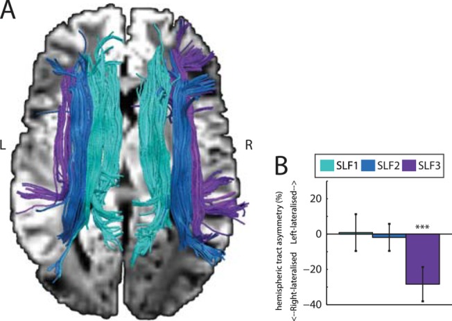
Fig 3. (A) Tractographic rendering of SLF branches in one subject obtained using diffusion MRI. The medial branch (SLF1) is shown in sky blue, the middle branch (SLF2) is shown in dark blue, and the lateral branch (SLF3) is shown in purple. These branches were identified by following the tracts intersecting coronal slices passing through both parietal cortex and, respectively, the superior frontal gyrus (SLF1), middle frontal gyrus (SLF2), and precentral gyrus (SLF3). (B) Group average hemispheric tract asymmetry for the three SLF branches. Consistent with previous work [21], only SLF3 shows consistent right lateralization (t(25) = -6.02, p < 0.0001). SLF1 and SLF2 are not lateralized (SLF1: t(25) = 0.17, p = 0.87. SLF2: t(25) = -0.51, p = 0.62). Error bars represent 95% confidence intervals. *** indicates p < 0.0001.

