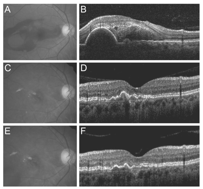
Fig. 1. Fundus photography and optical coherence tomography findings of an eye with submacular hemorrhage secondary to polypoidal choroidal vasculopathy. At the time of diagnosis, visual acuity was measured as 20 / 100 (A,B). At 6 months, the hemorrhage had resolved completely, and visual acuity had improved to 20 / 25 (C,D). The eye was treated with 5 ranibizumab injections during the 28-month follow-up period. At 28 months, visual acuity was maintained at 20 / 25 (E,F).

