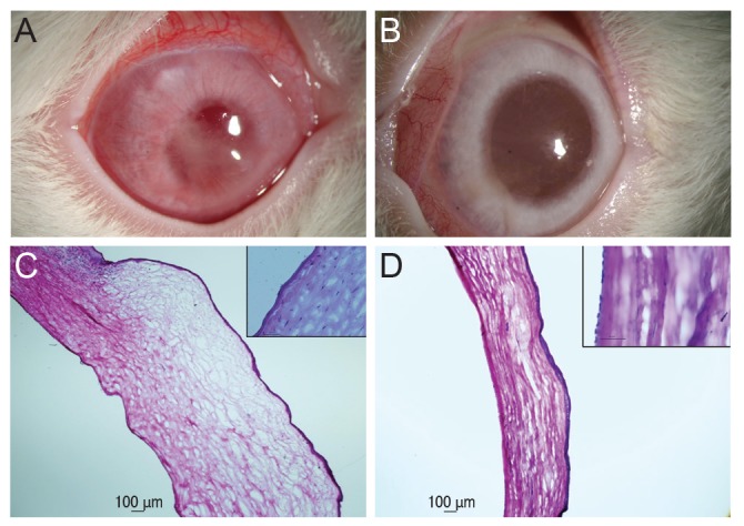
Fig. 1. (A) Slit lamp photograph of an eye 24 hours after injection of Cosopt. A rabbit eye in the Cosopt group showing severe corneal haze and conjunctival vascular injection. (B) Slit lamp photograph of an eye 24 hours after injection of Cosopt-S. A rabbit eye in the Cosopt-S group showing minimal corneal haze and conjunctival vascular injection, the extent of which was much more mild than that of Cosopt-treated eyes. (C) Histopathologic photomicrograph of a rabbit cornea 24 hours after injection of Cosopt. Cornea showing severe stromal edema. Many endothelial cells were lost (inset). (D) Histopathologic photomicrograph of a rabbit cornea 24 hours after injection of Cosopt-S. Significant stromal edema is absent. A single layer of endothelium is well observed (inset) (hematoxylin and eosin, ×40; inset ×400).

