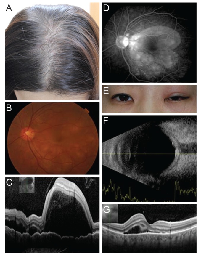
Fig. 1. (A) Alopecia developed several months before ophthalmic symptoms. (B) Initial fundus photography of the left eye, diffuse serous retinal detachment was observed. (C) Initial optical coherence tomographs showed subretinal fluid with serous retinal detachment, involing macula. (D) Initial fluorescein angiography showed some leak points and pooling into the subretinal space. (E) Recurred state, there was upper lid erythema, swelling and ptosis. (F) Recurred state, the ultrasonographs showed the increased thickness of the posterior scleral wall and the typical T sign caused by the spread of inflammation along the Tenon's space into the optic nerve sheath. (G) Recurred state, the serous retnal detachment was relapsed in optical coherence tomographs. But it was less severe than initial state.