
Fig. 2.
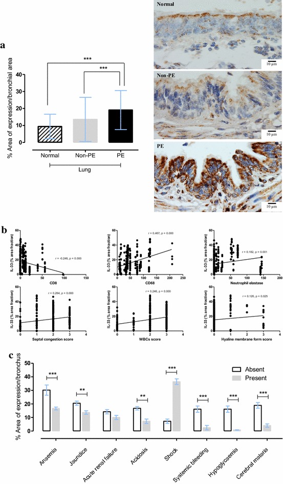
Immunohistochemical expression of bronchial IL-33 expression is increased in malaria cases with pulmonary oedem. a Bar chart comparing of bronchial IL-33 expression in normal, non-PE and PE lung samples and photomicrographs of immunohistochemical staining of bronchial IL-33 localization; b significance correlation plot of bronchial IL-33 expression to leukocyte count and histopathological score; c bar chart comparing of bronchial IL-33 expression to clinical manifestations