
Fig. 6.
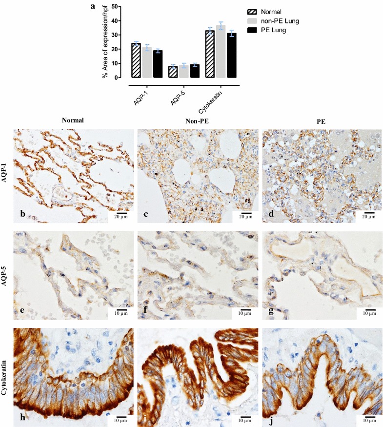
Immunohistochemical staining for AQP-1 and -5 and bronchial cytokeratin. a Bar chart comparing the degree of alveolar expression of AQP-1 and -5 and bronchial expression of cytokeratin in normal, non-PE, and PE-lung samples; photomicrographs of immunohistochemical staining of AQP-1 and -5 (localized on alveolar vessel and alveolar epithelium, respectively) and bronchial cytokeratin among normal (b, e, h), non-PE (c, f, i) and PE (d, g, j) lung samples