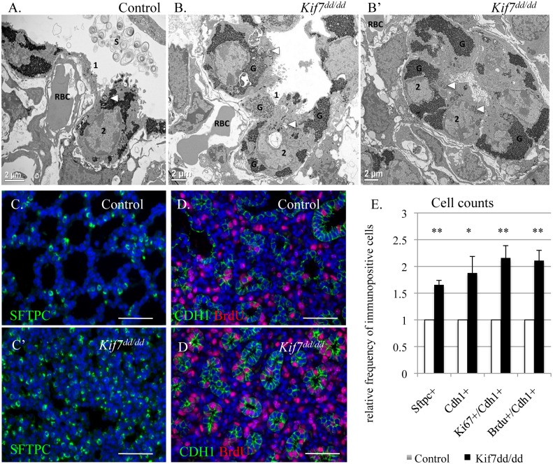
Fig 3. KIF7 is a negative regulator of cell proliferation within the respiratory epithelium.
(A.-B’) Transmission electron micrographs of E18.5 control and Kif7 dd/dd mutant respiratory airways. RBC (red blood cell) within the capillary adjacent to a type 1 AEC (1), AEC2 (2), Glycogen (G), Surfactant (S), Arrowheads denote lamellar bodies. (C.-C’) Immunofluorescent staining of SFTPC in E18.5 control and Kif7 dd/dd mutant lungs. (D.-D’) Co-immunoflorescent staining of Brdu (red) and CDH1 (green) in E17.5 control and Kif7 dd/dd mutant lungs. (E.) Quantification of relative number of immunopositive cells in control and Kif7 dd/dd mutant tissue sections. Immunopositive cells were counted and then divided by the total number of cells for control and mutant tissue sections. Control values were set as 1, and mutant values were divided by control values to provide a relative frequency of immunopositive cells per tissue section. At least 4 consecutive tissue sections were counted and averaged for an individual, from at least 4 sets of control and Kif7 dd/dd mutant lungs. * P<0.05, **P<0.01.

