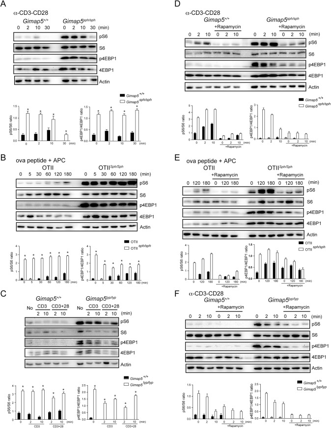
Fig 1. GIMAP5 regulates the activity of mTORC1.
(A and C) CD4+ T cells from control and Gimap5 deficient mice (A) and rats (C) were left un-stimulated or stimulated with 5 μg/μL anti-CD3 or anti-CD3/CD28 for the indicated period. Phosphorylation and the total amount of the indicated proteins in the whole cell lysates were evaluated by Western blot. (B) CD4+ T cells from the OTII TCR-transgenic control and Gimap5 sph/sph mice were co-cultured with irradiated APC pulsed with indicated period were lysed and analyzed by Western blot. (D and F) CD4+ T cells from control and Gimap5 deficient mice (D) and rats (F) were treated with vehicle or 200 nM rapamycin for 30 min followed by TCR cross-linking at different time points. Cell lysates were examined by Western blot using the indicated antibodies. (E) CD4+ T cells from the OTII Gimap5 sph/sph and control mice were pretreated with vehicle or 200 nM rapamycin for 30 min before simulation with ova peptide for the indicated duration. Cell lysates were probed with specific antibodies. (A-F) Representative data from 3 independent experiments are shown. Histograms show densitometric data from 3 experiments. * p<0.05 control vs mutant cells.

