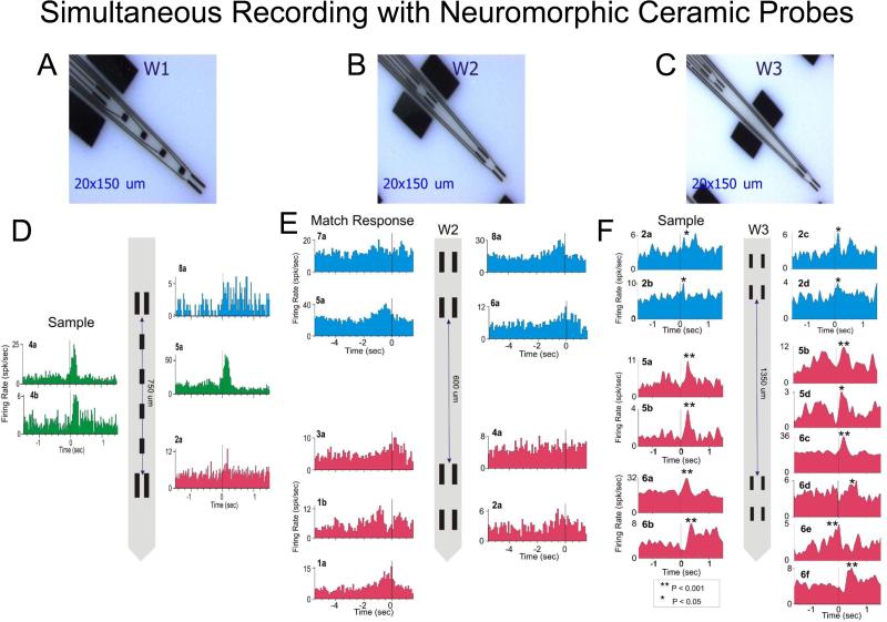
Figure 2. Example of simultaneous recordings of prefrontal neurons with neuromorphic multi-electrode arrays.
A,B,C. Illustration of configuration for three different types of neuromorphic probes W1, W2, W3 used in columnar recordings. D,E,F. Example of simultaneous recordings in the prefrontal cortex. The code color for the neural activity in cortical layers is: layer 2/3 (blue), layer 4 (green) & layer 5 (red). Peri-event histograms (PEHs) of cell activity simultaneously recorded with neuromorphic probes during a single session. Separation distance of the recording pads is shown for each MEA diagram with cells recorded from those locations indicated by different markers.

