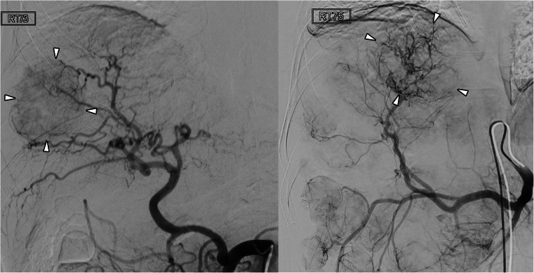
Fig. 2.
Celiac arteriogram during TACE in two patients with neuroendocrine tumour. Left panel was acquired on the preceding imaging platform and the right panel on the new imaging platform. Both arteriograms were of diagnostic quality, showing the tumour-feeding arteries and the tumour blush (arrowheads). However, the new imaging platform resulted in a significantly lower radiation exposure during the acquisition of the celiac arteriogram

