Figure 4.

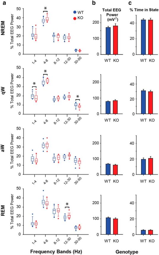
TASK channel deletion on cholinergic neurons alters electrocortical activity but not sleep–wake architecture. a, Percentage of total EEG power across sleep–wake states in the different frequency bands for ChAT-Cre:TASK+/+ (WT, blue) and ChAT-Cre:TASKf/f (KO, red) mice under baseline conditions of aCSF microperfusion into the basal forebrain (n = 20 WT and n = 20 ChAT-Cre:TASKf/f mice). In the box-and-whisker plots: Boxes represent the second and third quartiles separated by the median. Bottom and top whiskers represent the 10th and 90th percentiles, respectively. Individual dot symbols represent the extreme (i.e., highest and lowest) values from animals in each group. Groups were compared using a two-way repeated-measures ANOVA (factors being genotype and frequency band). b, Total EEG power (1–50 Hz) in the different sleep–wake states in WT and ChAT-Cre:TASKf/f mice. Values are mean ± SEM. Groups were compared using a two-way repeated-measures ANOVA (factors being genotype and state). c, Percentage of time spent in the different sleep–wake states in WT and ChAT-Cre:TASKf/f mice. Values are mean ± SEM. Groups were compared using a two-way repeated-measures ANOVA (factors being genotype and state). *p < 0.05, significant difference between groups. For further details, see Results.
