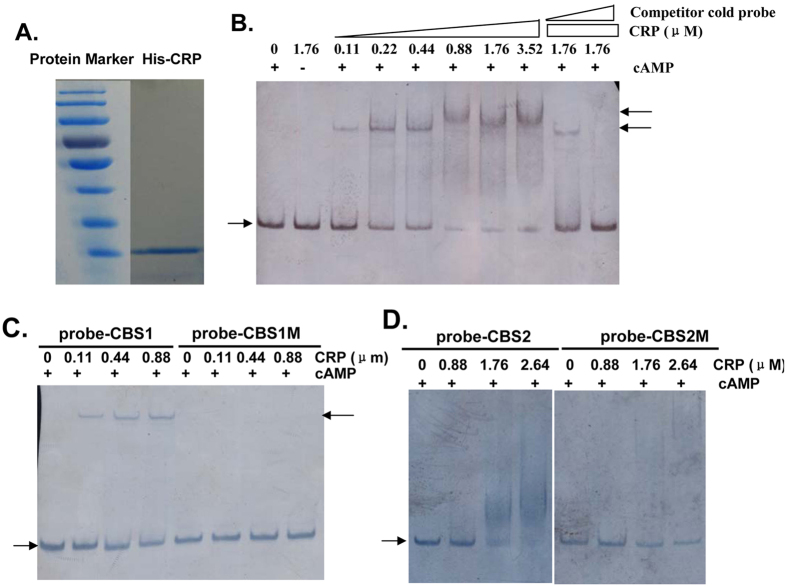
Figure 6. Binding of the cAMP-CRP complex to the promoter region of tfoXVC.
EMSA were performed as described in the Methods section to determine whether there was a direct interaction between cAMP-CRP and the promoter region of tfoXVC. The arrow on the left side indicates the unbound free probe, whereas the arrow on the right side indicates the probe bound with CRP. (A) The purity of His-CRP analyzed with SDS-PAGE. (B) A biotin-labeled 152-bp DNA probe containing two CRP binding sites (10 ng) was incubated with increasing amounts of CRP in the presence of cAMP (0.1 mM). For the competition analysis, the same, but unlabeled, 152-bp DNA was included as 100-fold and 300-fold concentrations relative to the labeled probes. (C) The biotin-labeled 75-bp DNA fragments containing the original distal CRP binding site (probe-CBS1) or the corresponding mutagenized binding site (probe-CBS1M) were used as probes in a gel shift assay. (D) The biotin-labeled 97-bp DNA fragments containing the original proximal CRP binding site (probe-CBS2) or the corresponding mutagenized binding site (probe-CBS2M) were used as probes in the gel shift assay.

