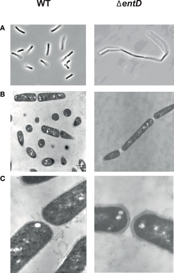
Figure 2.

Microscopic observation of cells in exponential growth phase cultures of B. cereus wild-type and ΔentD strains. (A) Phase contrast microscopy × 1000. (B) Thin-layer electron microscopy × 7000. (C) Thin-layer electron microscopy × 35,000. Cultures were grown in MOD medium supplemented with 30 mM glucose under aerobiosis. Samples were harvested during exponential growth phase. Photographs shown in the figure are of representative samples from three independently grown cultures.
