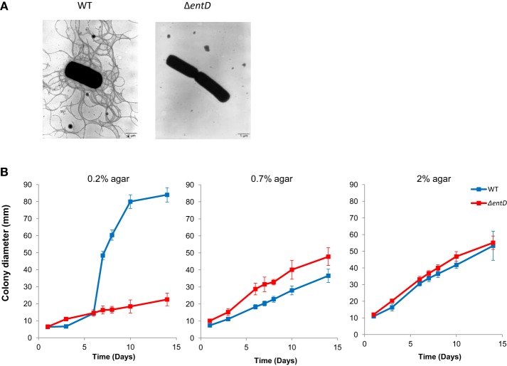
Figure 7.
Flagellation and motility of B. cereus wild-type and ΔentD mutant strains. (A) Negative staining electron micrographs (× 7000) of wild-type and ΔentD mutant strains. (B) Motility of wild-type and ΔentD mutant strains. Diameters of motility haloes were measured during 2 weeks on TrB agar plates containing 0.2, 0.7, and 2% agar, respectively. The data shown are the mean ± standard deviations of triplicates. Statistical differences between WT and mutant strains were evaluated with the Student's t-test.

