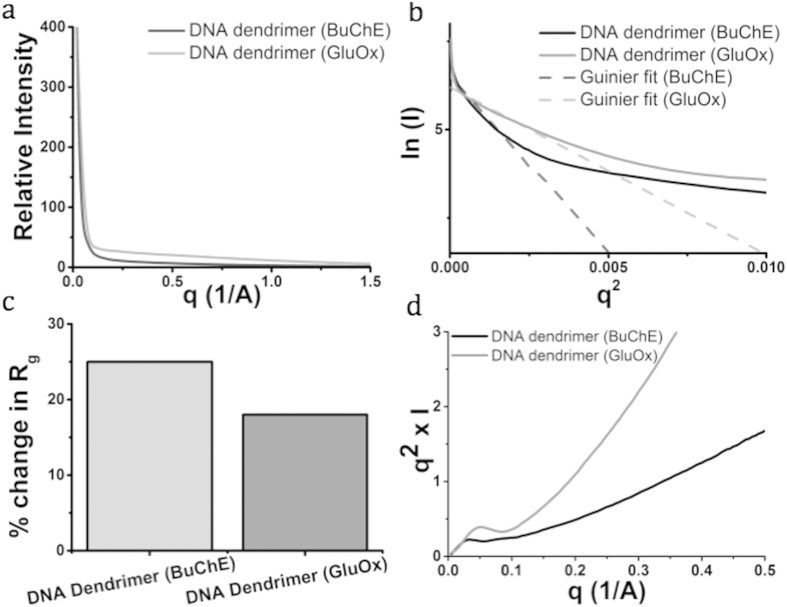
Figure 2. Small angle x-ray scattering (SAXS) of DNA dendrimers with butyrylcholinesterase (BuChE) or glucose oxidase (GluOx) attached was done to further assess the structure and stability of the DNA dendrimers.
We performed SAXS with two samples of DNA dendrimers (at 1 mg/mL in PBS). (a) SAXS scattering data depicting the standard plots of circularly averaged intensity as a function of q, where q = 4π/λ[sin(θ/2)] (θ = scattering angle). (b) Guinier plot of scattering data demonstrates the conformational flexibility of the dendrimer structures and yields an approximate size based on the Guinier slopes. (c) Quantified percent change in the radius of gyration (Rg), measured by the change in the Rg of the experimentally-determined value from the theoretical value. (d) Kratky plot of scattering data indicates the heterogeneous internal substructure of the DNA dendrimers.

