
Figure 3.
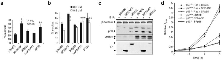
SF2/ASF overexpression protects E1A-sensitized MEF cells against apoptosis and enhances the proliferation of Ras-transformed cells. Primary wild-type MEF cells were transduced with retroviruses expressing the indicated human splicing factor cDNAs, or with the empty vector; some cells were cotransduced with adenovirus E1A or activated Ras, as indicated. Error bars indicate s.d. (a) After drug selection, cells coinfected with E1A were plated and serum-starved, and cell death was measured by trypan blue staining. P-values in pairwise comparisons to the pBABE control: *P = 5× 10⁻⁸; **P = 5 × 10⁻⁵; n = 3. (b) Cells coinfected with E1A were plated and treated with the indicated adriamycin concentrations after 24 h, and cell death was measured as in a. *P = 0.003, **P = 0.01, ***P = 0.006; n = 3; for SF2/ASF at low adriamycin, n = 1. (c) MEF cells were infected with the indicated retroviruses, plated after single or double selection (106 cells per 10-cm plate) and lysed in SDS after 24 h. Western blots were carried out using the indicated primary antibodies. The reduction in SRp55 in the presence of E1A was not observed with other cell lines (data not shown). (d) p53−/− MEF cells were transduced with the empty vector or with retroviruses expressing SF2/ASF or SRp55, either alone or with oncogenic Ras. Cells were fixed at 48-h intervals and stained with methylene blue. Each point represents the mean relative absorbance from six wells.