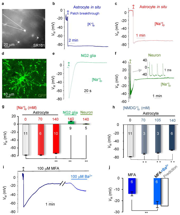
Figure 1. Physiological VM can be well-maintained in astrocyte recordings with reduced or K+ free pipette solutions in situ.
(A) SR101 staining of astrocytes in CA1 region. (B and C) Astrocyte VM recordings, first in cell-attached, then in whole-cell mode after membrane patch breakthrough (arrows). The resting VM’s were comparable between K+]p and [Na+]p solutions. (D and E) An NG2 glia identified from PDGF-driven GFP transgenic mouse CA1 region in situ, and its VM recording with [Na+]p. The VM showed an initial hyperpolarization and a following depolarization. (F) The VM recording from an interneuron with [Na+]p, showing an initially hyperpolarization (−40 mV), followed by a depolarization (~0 mV). A burst of spikes appeared shortly after patch breakthrough (upper inset). (G) Summary of the VM values for the cell types and conditions indicated (one-way ANOVA with post-hoc F test). (H) Summary of the VM values from astrocytes recorded with various pipette NMDG+ concentrations in situ (one-way ANOVA with post-hoc F test). (I) Gap junction inhibition by 100 μM MFA depolarized VM and the subsequent Kir4.1 inhibition (100 μM Ba2+) hyperpolarized VM as predicted by the mathematical model (Supp. Info. Fig. 4). (J) Summary of the VM values in MFA, MFA+Ba2+, and a predicted value for the latter from the model (paired sample t test). *P < 0.05.

