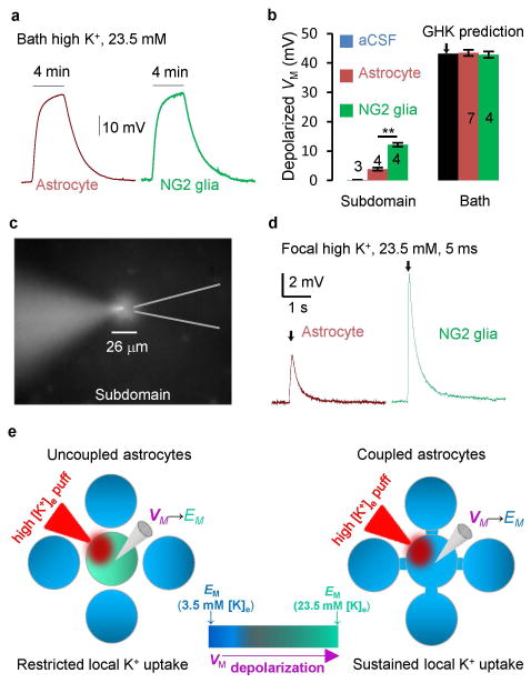
Figure 5. Syncytial coupling attenuates local elevated extracellular K+-induced VM depolarization.
(A) Bath 23.5 mM high K+-induced VM depolarization from an astrocyte and an NG2 glia, as indicated. (B) Summary of bath and local high [K+]e induced VM depolarization. (C) A 5 ms local high [K+]e was applied to a recorded astrocyte with an affected subdomain area monitored by SR101 included in the high [K+]e solution (two sample t test). (D) A 5 ms local high [K+]e puff induced VM depolarization in an astrocyte and an NG2 glia. (E) Schematic illustration of VM response to a high [K+]e puff. In an uncoupled astrocyte, the VM quickly depolarizes to the new elevated [K+]e established EM. Once VM → EM, the K+ uptake stops. In coupled astrocytes, the VM remains close to a constant. This maintains the driving force, VM − EM, for a “sustained K+ uptake”. *P < 0.05, one way ANOVA test for comparison of the VM depolarization between astrocytes and NG2 glia.

