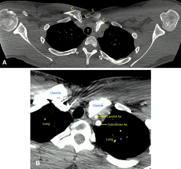
Figure 2.
A‐2B. An axial view of a CT scan (Figure 2A), revealing a posterior dislocation of the left sternoclavicular joint. Note the posterior position of the medial head of the involved clavicle (C) compared to the uninvolved clavicle (C) in relationship to the sternum (S). Figure 2B, an axial view of a CT scan with injected contrast (angiography), shows the proximity of the dislocated clavicle (C) to the trachea (T). Note the dislocated clavicle sitting directly over the left carotid and subclavian arteries.

