
Figure 3. Brichos interacts with fibrillar but not monomeric Aβ42.
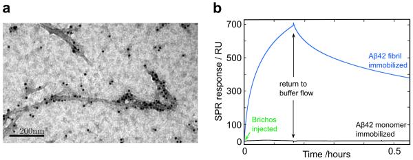
(a) Images using TEM with a nano-gold conjugated secondary antibody against anti-Brichos antibodies show that the chaperone binds to Aβ42 fibrils. (b) SPR analysis verifies the specific binding to fibrils and allows determination of the association (kon ≈ 5.1 × 10³ M−1s−1) and dissociation (koff ≈ 2.1 × 10⁻⁴ s−1) rate constants, implying an apparent equilibrium dissociation constant KD ≈ 40 nM. No binding was observed to the monomer or to control in the absence of Aβ42.