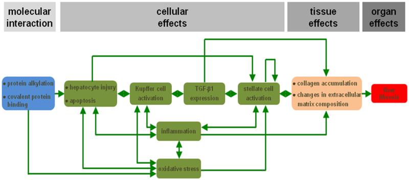
Figure 1. AOP for drug-induced liver fibrosis.
The MIE (blue) is considered protein alkylation and covalent protein binding in the liver. This serves as a trigger to provoke hepatocyte injury, including apoptosis, which in turn activates Kupffer cells. As a result, transforming growth factor beta 1 (TGF-β1) expression is induced, which is a key factor for stellate cell activation. The latter goes hand in hand with the occurrence of inflammation and oxidative stress. The different KEs at the cellular level (green) are interconnected in several ways. The overall end result is accumulation of collagen and changes in the extracellular matrix composition in the liver (orange), which becomes clinically manifested as the adverse outcome, namely liver fibrosis (red) (reproduced with permission from20).

