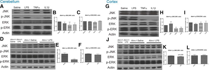
Figure 6.

MAP kinase levels in the cerebellum and frontal cortex after different inflammatory challenges. In cerebellum, phosphorylation levels of JNK trended lower after inflammatory stimuli (A, B). This effect was enhanced in Atm −/−, where LPS significantly reduced the levels of phospho-JNK (D, E). In cortex, the reverse situation was found. Phospho-JNK decreased significantly in wild-type after immune challenge (G, H), whereas in Atm −/− phospho-JNK only trended lower after LPS. In both regions and both genotypes, the levels of ERK phosphorylation were largely unchanged after an immune stimulus (C, F, I, L). n = 5 for wild-type animals and n = 3 for Atm−/− animals.
