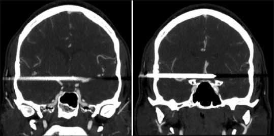
Figure 3.

Sections of a computed tomography angiogram are confirming intact internal carotids. The right M1 to the proximal M3 middle cerebral artery and anterior communicating artery regions are not visualized. However, there is normal distal filling of the right middle cerebral artery beyond the beam-hardening artifact, with no obvious vessel perforation, dissection, thrombosis, or branch occlusion
