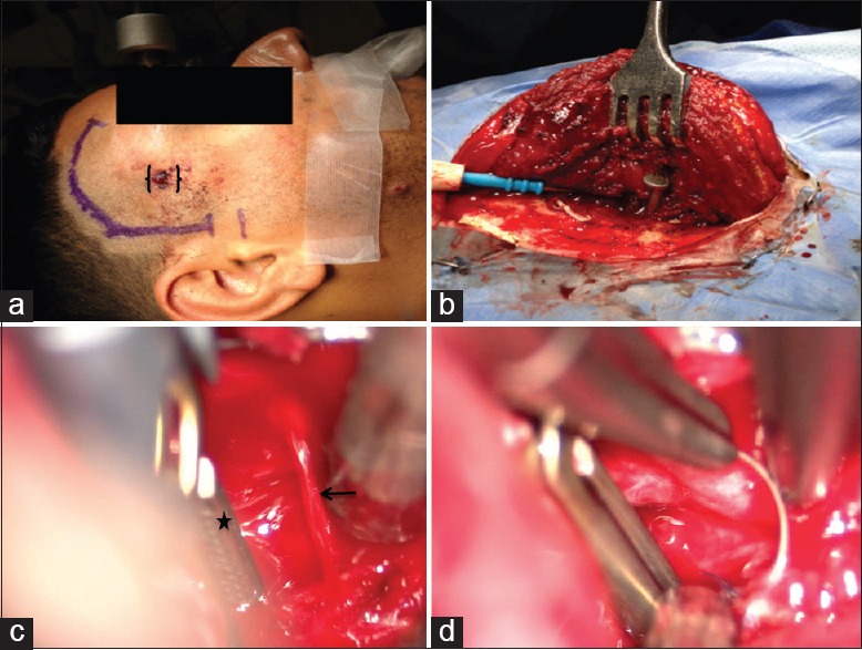
Figure 4.

Intraoperative images demonstrating the entry point of the nail (double brackets), proposed pterional incision (a) and temporalis reflection (b). A jet of blood (arrow) exsanguinating from the M1 vessel and application of a proximal temporary aneurysm clip (star) (c). Repair of the arteriotomy (d)
