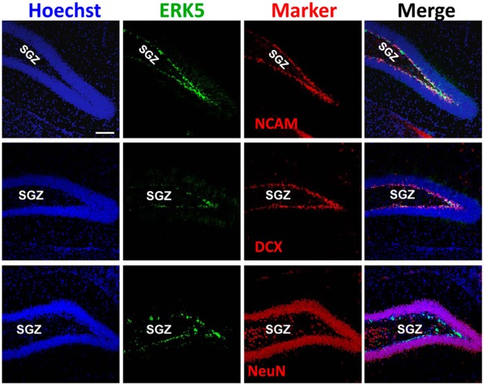
Figure 2.
ERK5 MAPK expression in the dentate gyrus of the adult mouse brain. Images are representative immunostaining of coronal sections of adult mouse brain tissue showing ERK5 protein expression (green) primarily in transiently amplifying progenitors and/or newborn neurons (doublecortin+ (DCX) or NCAM+) but not in mature neurons (NeuN, red) in the SGZ. Hoechst staining (blue) was used to identify all cell nuclei. Scale bar represents 100 μm and applies to all panels.

