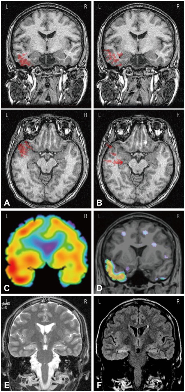
Fig. 1.

EEG and MEG dipole source analysis of patient #14 who had left temporal lobe epilepsy. A: EEG dipoles [high-pass filter (HPF)=3 Hz, goodness of fit (GOF) levels ≥70%] were localized in left basal and anterior temporal regions. B: MEG dipoles (HPF=3 Hz, GOF levels ≥70%) were localized in left anterior to middle temporal regions. C: Ictal SPECT showed left temporal hyperperfusion. D: SISCOM showed left anterior to mid temporal hyperperfusion. (E) T2-weighted MRI and (F) FLAIR MRI showed left hippocampal sclerosis. L: left, R: right.
