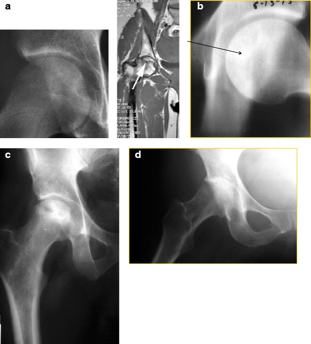
Fig. 1.

Plain radiographs showing different stages of disease. a Ficat Stage I ON patient with normal radiograph and abnormal MRI. b Ficat stage II ON patient with arrow pointing to sclerotic lesion. c Ficat stage III ON patient with evident femoral head collapse. d Ficat stage IV ON patient with acetabular involvement
