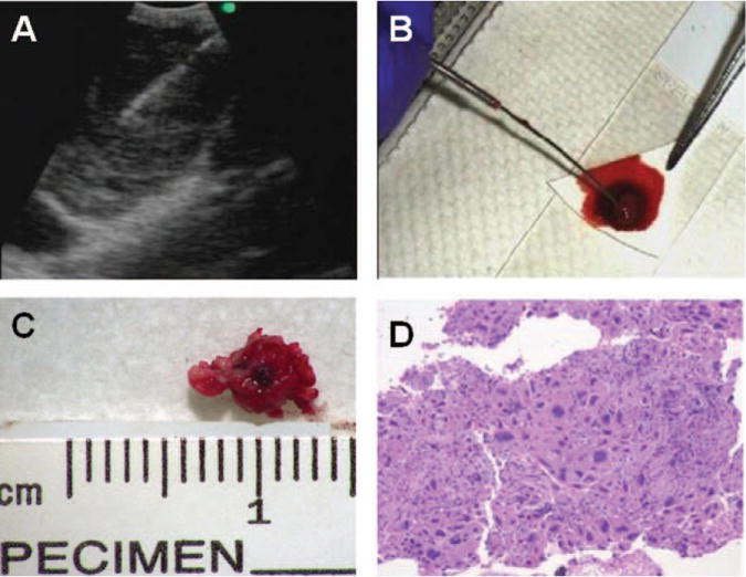
FIGURE 1.

Cell block preparation using the tissue coagulum clot (TCC-CB) method is shown. (A) Endobronchial ultrasound imaging of the station 7 lymph node with a metastatic, poorly differentiated adenocarcinoma is shown. (B) A TCC-CB is shown on the filter paper. (C) The actual size of the TCC-CB is shown. (D) Hematoxylin and eosin staining of the TCC-CB section from the same lymph node with a metastatic, poorly differentiated adenocarcinoma as shown in Panels A through C is shown (× 200).
