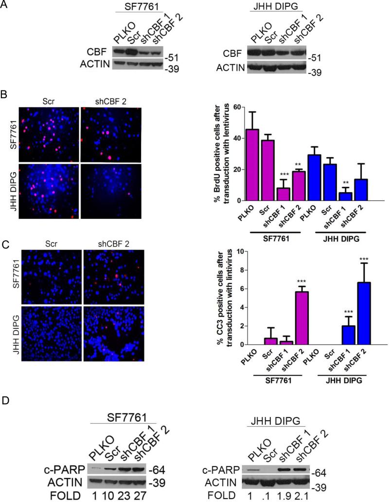
Figure 4.
Core binding factor (CBF) shRNA phenocopies γ-secretase inhibitor (GSI) in diffuse intrinsic pontine glioma (DIPG). (A) Western blot showing decreased CBF1 expression with SF7761 (left) and JHH-DIPG1 (right) cells after infection with lentiviral vectors for shCBF. (B) Left, representative anti-bromodeoxyuridine (BrdU) immunofluorescence (red) showing decreased proliferation in cells transduced with lentiviral vectors for shCBF compared to scramble (Scr). DAPI (blue) counterstains nuclei. Magnification: 400X. Right, quantification of percent BrdU positivity in SF7761 and JHH DIPG1 transduced with lentiviral vectors for shCBF (right) **p < 0.01; ***p < 0.001 by ANOVA. (C) Left, representative anti-cleaved caspase 3 (CC3) immunofluorescence (red) showing increased apoptosis in cells transduced with lentiviral vectors for shCBF compared to Scr control. DAPI (blue) counterstains nuclei. Magnification: 400X. Right, quantification of percent CC3 positivity in SF7761 and JHH DIPG1 transduced with lentiviral vectors for shCBF. **p < 0.01; ***p < 0.001 by ANOVA. (D) Western blot showing increased cleaved PARP expression with SF7761 (left) and JHH-DIPG1 (right) cells transduced with lentiviral vectors for shCBF. Numbers below the blot represent fold change compared to pLKO control.

