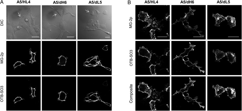
Fig. 7.
Determining biosensor activities of tandem scFv dimers when fused to a cell surface receptor. (A) Micrographs of mammalian cells expressing different tandem scFvs fused to ADRB2. Images were acquired using two different emission channels with corresponding excitation laser wavelengths. The white arrows on the DIC images indicate untransfected cells. (B) Same as A; however, the cells were initially labeled with MG-2p fluorogen, washed and incubated with an ADRB2 agonist, and then washed and replaced with medium containing only OTB-SO3 fluorogen. The MG-2p emission channel shows the ADRB2 agonist response of intracellular vesicular traffic. The OTB-SO3 emission channel only shows ADRB2 cell surface signal due to the removal of agonist from the medium. All the experiments were performed using 100 nM MG-2p and OTB-SO3 fluorogens, and the scale bar represents 30 μm.

