Fig. 5.

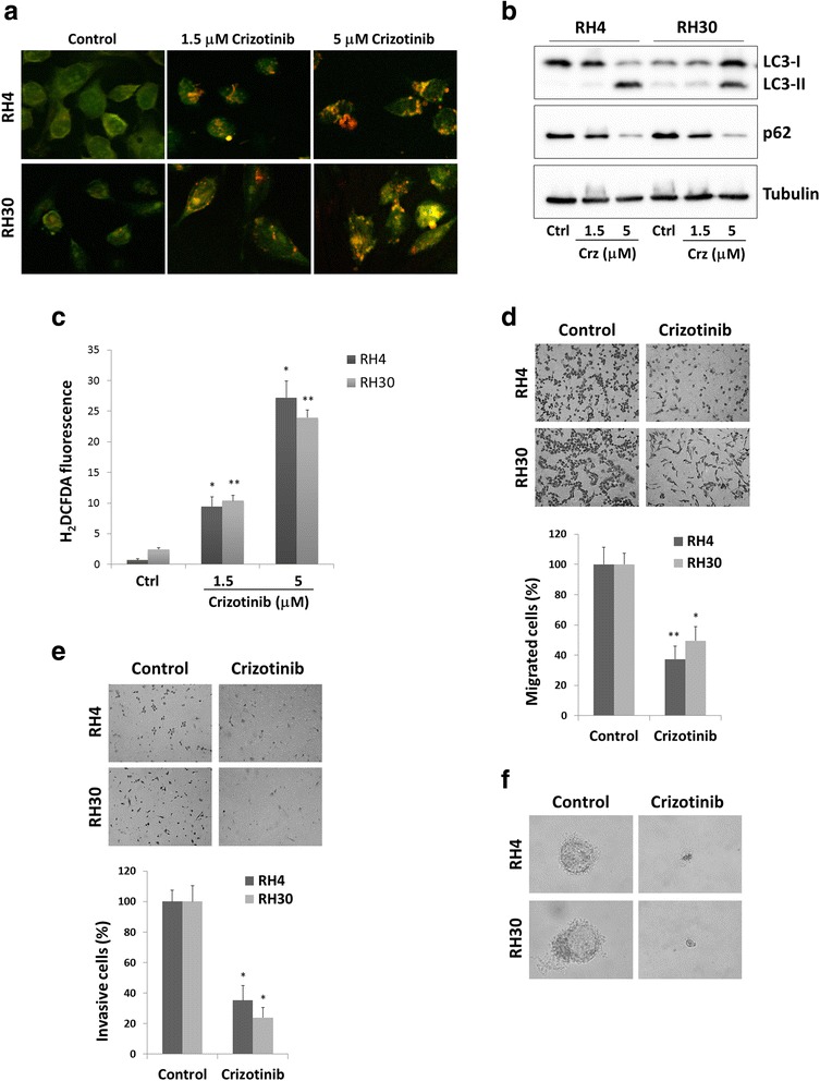
Effects of crizotinib treatment on autophagy, migration, invasion and colony formation in RH4 and RH30 cell lines. a Acridine orange staining showed that RH4 and RH30 DMSO-treated control cells exhibited no autophagosomes, whereas crizotinib-treated cells showed abundant cytoplasmic acidic vesicular organelles (AVO) formation, a characteristic of autophagy. Samples were observed under an ApoTome fluorescence microscope. b LC3 and p62 protein levels were examined in RH4 and RH30 cells treated with crizotinib (Crz) at various concentrations for 24 h. Control cells (Ctrl) received DMSO vehicle. c ARMS cells were treated with crizotinib (1.5 and 5 μM) for 24 h and H2DCFDA was added 45 min before collecting cells. H2DCFDA fluorescent intensities were analysed by FACSCalibur. Control cells (Ctrl) with H2DCDA staining were used as a negative control. Bar charts represent the mean fluorescence ± SD for each sample (*, p-value <0.05 compared with the respective negative control; **, p < 0.01). Three independent experiments were performed. d Crizotinib (1.5 μM) treatment led to a decreased cell migration in both RH4 and RH30 cells. Representative images of migrated cells using the transwell migration assay (crystal violet staining, magnification of 20×). Histograms represent the average values ± SD as determined by counting 8 fields of cells under the microscope. Three independent experiments were performed, each in triplicate (*, p-value <0.05 compared with the respective negative control; **, p < 0.01). Control cells were treated with DMSO. e Crizotinib (1.5 μM) treatment led to decreased cell invasion in both RH4 and RH30 cells. Control cells were treated with DMSO alone. Representative images of invasive cells using the Matrigel-invasion assay (crystal violet staining, 20× magnification). Histograms represent the mean value ± SD as determined by counting 8 fields of cells under the microscope. Three independent experiments were performed, each in triplicate (*, p < 0.05). f Crizotinib (1.5 μM) induction led to a decreased cell colony formation capacity in both RH4 and RH30 cells. Control cells were treated with DMSO alone. Representative images of a single cell derived-sphere in mocked control and treated cells cultured in soft agar for 21 days (original 40× magnification)
