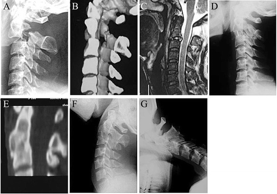
Fig. 3.

Images of a 40-year-old male patient. a and b, preoperative lateral X-ray and CT scans showing a type II Hangman’s fracture with severe translation. c, MRI with sagittal section showing C2/3 intervertebral disc injury. d, 3-month postoperative lateral X-ray showing adequate reduction and bony fusion. e, CT with sagittal reconstruction showing solid fusion and fracture healing. f and g, 24-month flexion/extension lateral X-rays showing no range of motion at the fusion site
