Fig. 4.

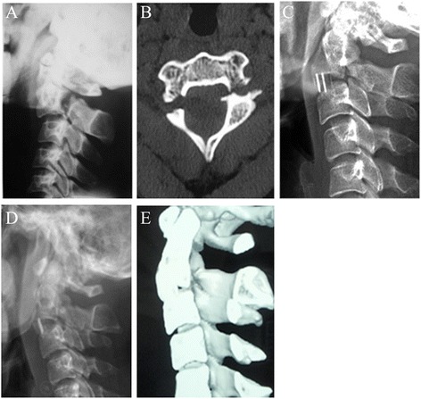
Images of a 26-year-old female patient. a and b, preoperative lateral X-ray and CT scans showing a type II hangman’s fracture with severe translation. c, Postoperative lateral X-ray showing minor implant migration at 1 week postoperatively. d, The X-ray showed the implant remained the same position during follow-up. e, Bone fusion was confirmed using CT scan at 4 months postoperatively
