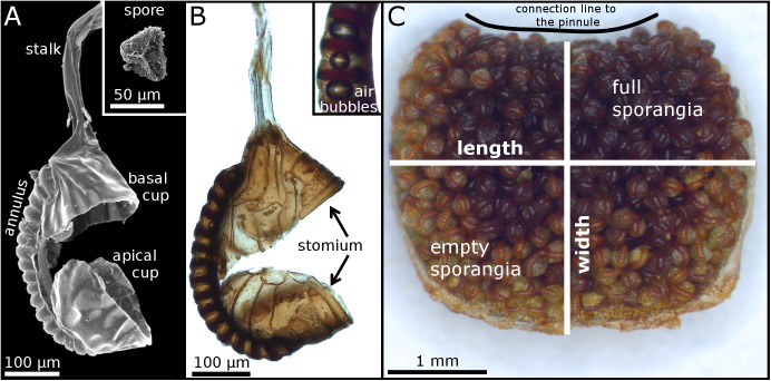
Fig 2. Leptosporangia on the underside of a false indusium of A. peruvianum.
(A) Scanning electron microscopy (SEM) image of a ruptured empty sporangium. The inset shows a trilete spore. (B) Light microscopy (LM) image of a ruptured empty sporangium. The inset shows annular cells with gas bubbles that occurred due to cavitation inside the cells. (C) Underside of a detached false indusium with sporangia that partly have already shed their spores. The connection line to the pinnule margin is indicated.

