
Figure 5.
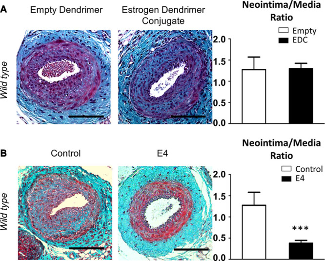
Genomic functions of estrogen receptor α (ERα) mediate protection from intimal hyperplasia. A, Four-week-old wild-type female mice were ovariectomized and subcutaneously implanted with estrogen dendrimer conjugate or empty dendrimer-eluting osmotic minipumps. Two weeks later, animals were submitted to mechanical injury of the femoral artery. Arteries were harvested 28 days after the injury for morphometric analysis. Left, Representative images of cross sections of femoral arteries of indicated mice stained with Masson Trichrome. Bars, 100 µm. Right, Quantitative analysis of neointima/media ratio of indicated mice. Values are presented as mean±SEM (n=10–12 mice per group), and statistically compared with Mann–Whitney U test. B, Four-week-old wild-type female mice were ovariectomized and subcutaneously implanted with control of estetrol (E4)-eluting osmotic minipumps. Two weeks later, animals were submitted to mechanical injury of the femoral artery. Arteries were harvested 28 days after the injury for morphometric analysis. Left, Representative images of cross sections of femoral arteries of indicated mice stained with Masson Trichrome. Bars, 100 µm. Right, Quantitative analysis of neointima/media ratio of indicated mice. Values are presented as mean±SEM (n=7–12 mice per group), and statistically compared with Mann–Whitney U test. ***P<0.001.