
Fig 1. Experimental set-up.
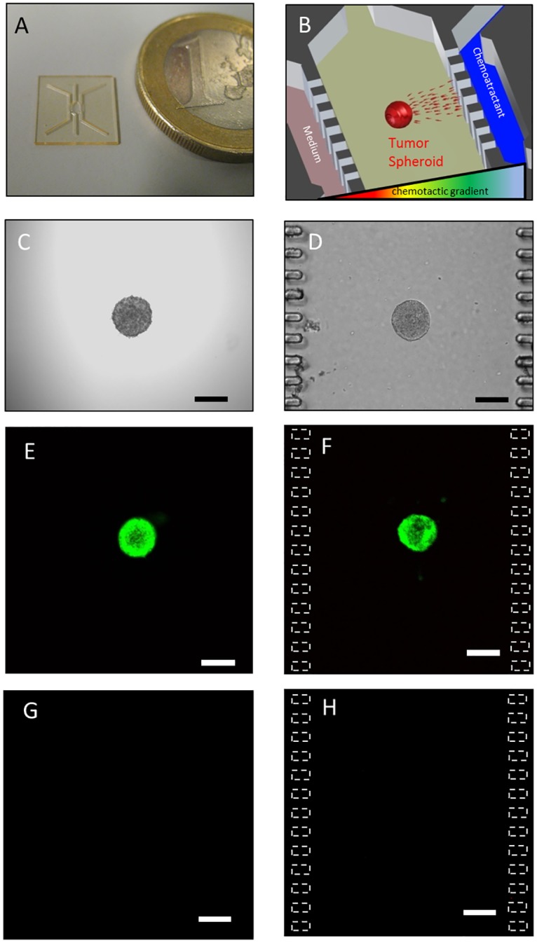
(A) SU–8 fabricated microdevice. (B) Experimental scheme. (C) OSC–19 spheroid of 2000 cells in the hanging drop. (D) Same spheroid after embedding in 1.5 mg/ml collagen hydrogel within the microdevice. Treatment of the spheroids with FDA/PI after 48 hours in a hydrogel in a 96-well plate or within the microdevice shows the spheroid is intensely green (E and F respectively) whereas less than five red cells where observed (G and H respectively), Z projection of the whole spheroid is shown. Scale bar is 200 μm.