
Fig 7. Chemotactic behavior of OSC–19 individual cells.
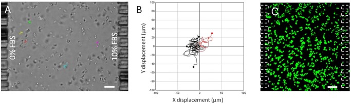
(A) Individual OSC–19 cells were embedded in collagen hydrogel within the central microchamber. Media supplemented with 10%FBS was perfused through one lateral microchannel, whereas basal media was used in the other. Tracks of migrating cells are shown. (B) Individual cell trajectories are plotted, showing those with a net displacement to the right in red, and those displaced to the left in black. (C) Isolated OSC–19 cell viability after 30 hours under gradient conditions, viable cells are shown in green. Scale bar is 100 μm.