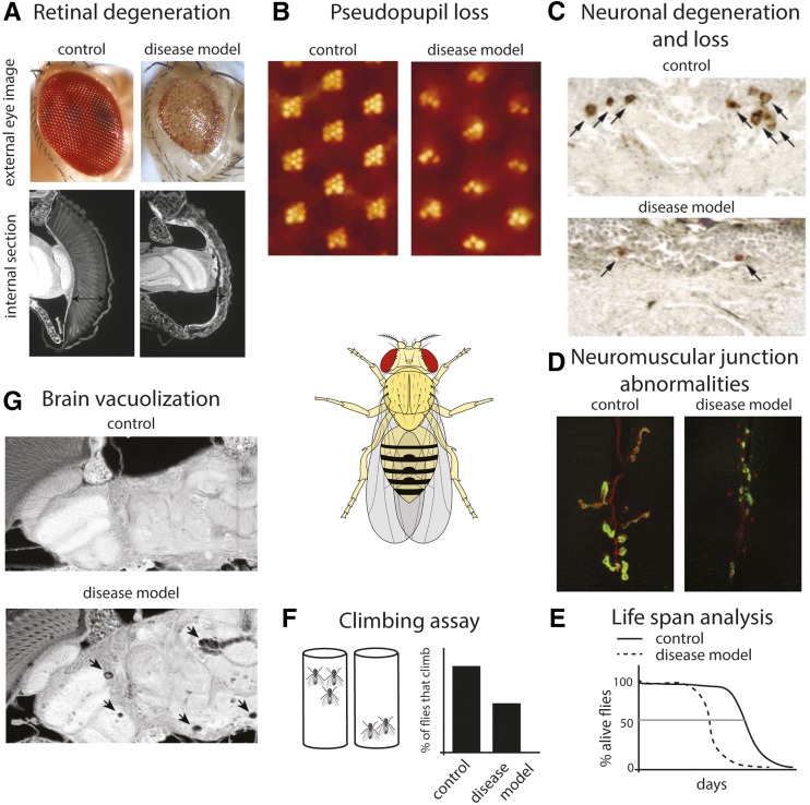
Figure 2.
Examples of robust assays to assess neural degeneration and dysfunction in Drosophila. We illustrate assays that can be used in disease models for a neurodegenerative effect. We note that similar approaches, but geared toward the features of the disease of interest, can be used in the characterization of fly models for other classes of human disease. (A) Using an eye-specific GAL4 driver, both the disease gene and candidate modifiers can be expressed in the eye. As the eye is a nonvital tissue, the effects of highly toxic genes can be assessed in adult flies without concerns of lethality. For large-scale screens, the external eye (top panel) offers a rapid readout. Compared with the normal eye, the degenerative eye can show disruption of ommatidial structure, reduced size, and loss of pigmentation. Internal sections (bottom panel) allow examination of the retinal tissue. The degenerative eye shows collapse of the retina (arrows). (B) The pseudopupil assay allows a quantitative measure of degeneration in adult flies by assessing the structure of the photoreceptor cells by counting the number of intact rhabdomeres (the specialized organelles of the photoreceptor cells). Each ommatidium is composed of eight photoreceptor cells. Using back illumination of the head, seven rhadomeres of each ommaditium can be visualized (left panel) and their structural deterioration over time due to degeneration can be monitored (right panel). (C) Labeling of specific neuronal cell types, either by using selective antibodies (such as antityrosine hydroxylase to label dopaminergic neurons) or by expression of markers using the GAL4-UAS system can allow one to assess the integrity of neurons in the brain [reprinted from Auluck et al. (2002)]. (D) Expression of disease genes in motor neurons, followed by immunostaining for presynaptic (red, anti-HRP) and postsynaptic (green, anti-Dlg1) neuromuscular junction (NMJ) markers can reveal NMJ abnormalities such as reduced bouton numbers and increase in “ghost boutons” that lack opposing postsynaptic structure (Kim et al. 2013b). Images are courtesy of N. C. Kim and J. P. Taylor (St. Judes Medical Center). (E) Lifespan analysis of flies expressing disease-related genes. Transgene expression can be limited to the nervous system, to specific neuronal cell types, or to the adult stage to bypass developmental effects by using the range of driver lines available. (F) The negative geotaxis climbing reflex of Drosophila can be used to examine motor deficits with age. Flies are tapped to the bottom of an empty vial and the number of flies that can climb above a certain height is recorded. (G) Brain degeneration in Drosophila is often accompanied by formation of vacuoles as shown here upon expression of a CAG-repeat RNA [reprinted from Li et al. (2008)]. Image of Drosophila in center, is courtesy of wiki commons.

