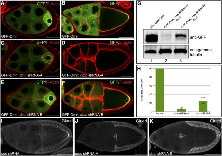
Figure 1.
shRNA-mediated depletion of Dmn. (A and B) Egg chambers expressing GFP-Dmn under the control of a maternal tubulin promoter were fixed and processed for immunofluorescence using an antibody against GFP (green). The egg chambers were also counterstained for F-actin (red). Representative stage 5 (A) and stage 10 (B) egg chambers are shown. (C and D) Egg chambers coexpressing dmn shRNA-A and GFP-Dmn were fixed and processed for immunofluorescence using an antibody against GFP. Representative stage 5 (C) and stage 10 (D) egg chambers are shown. (E and F) Egg chambers coexpressing dmn shRNA-B and GFP-Dmn were fixed and processed for immunofluorescence using an antibody against GFP. Representative stage 5 (E) and stage 10 (F) egg chambers are shown. dmn shRNA-A and dmn shRNA-B were expressed using a maternal α-tubulin-Gal4 driver (see Materials and Methods for details). GFP-Dmn was expressed using a construct in which the maternal tubulin promoter was cloned upstream of the GFP-dmn-coding sequence (Januschke et al. 2002). (G) Ovarian lysates were prepared from strains expressing GFP-Dmn (lane 1), from strains coexpressing dmn shRNA-A and GFP-Dmn (lane 2), or from dmn shRNA-B and GFP-Dmn (lane 3). The lysates were run on an SDS-PAGE gel and analyzed by Western blotting using an antibody against GFP (top). The same blot was then probed using an antibody against γ-tubulin (bottom). The level of γ-tubulin serves as a loading control. The images were captured digitally using a UVP bioimaging system. (H) Western blots from three separate experiments depicted in G were quantified using the VisionWorks software (UVP). The level of GFP-Dmn in strains coexpressing dmn shRNA-A or dmn shRNA-B were compared to the level of GFP-Dmn in the control strain. The error bars indicate standard deviation. ***P = 0.0001. (I–K) Egg chambers expressing a control shRNA against eb1 (H), dmn shRNA-A (I), or dmn shRNA-B (J) were fixed and processed for immunofluorescence using an antibody against Glued. The shRNAs were expressed using a maternal α-tubulin-Gal4 driver. Bar: A, C, and E = 20 μm; B, D, F, I, J, and K = 50 μm.

