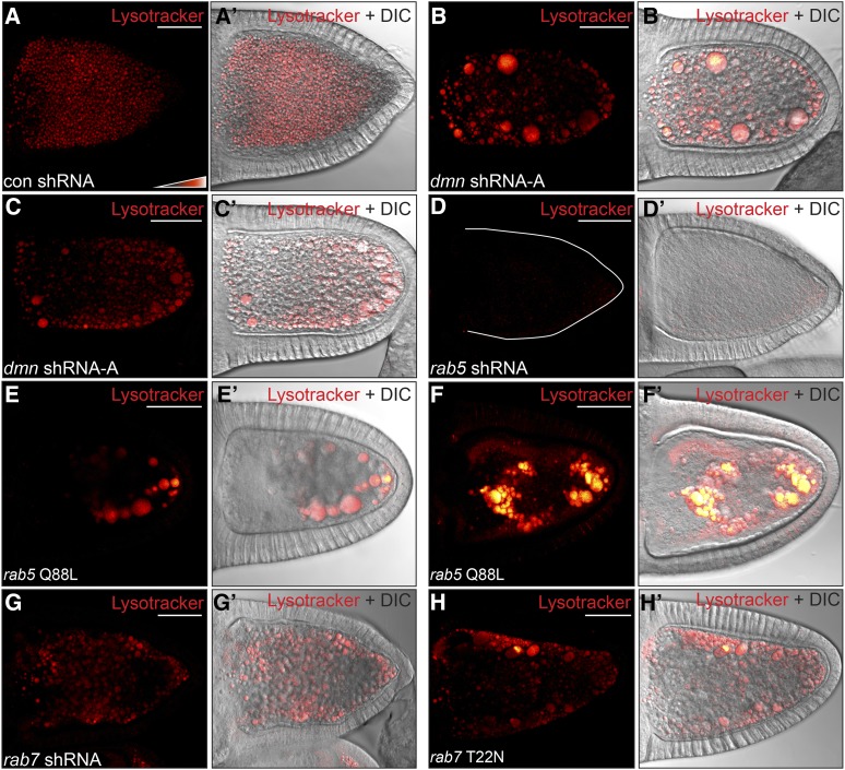
Figure 6.
The enlarged vesicles are Lysotracker-positive. (A–D) Egg chambers expressing a control shRNA (A), dmn shRNA-A (B and C) or rab5 shRNA (D) were processed live for Lysotracker staining. The egg chambers were then fixed and imaged. Lysotracker signal is displayed using a color-coded range indicator. Black pixels represent no signal, red pixels represent moderate levels of signal, and white pixels represent high signal. (E and F) Egg chambers expressing rab5 Q88L using the maternal α-tubulin-Gal4 driver were processed as in A. Approximately 50% of these egg chambers contained enlarged endosomes that displayed a similar level of Lysotracker staining as control oocytes (E and E′). The remainder contained enlarged endosomes that displayed a very high level of Lysotracker staining (F and F′). (G and H) Egg chambers expressing rab7 shRNA (G) or rab7 T22N (H) using the maternal α-tubulin-Gal4 driver were processed as in A. Depletion of Rab7 (G and G′) or overexpression of dominant negative Rab7 (H and H′) produced Lysotracker-positive enlarged endosomes.

